

Endoscopic Management of Lower Ureteric Stones Using an 8.5 Fr Pediatric Cystoscope: A Case-Based Technical Overview
Treating ureteric stones in toddlers is often more challenging than treating the same condition in adults. The smaller size of the urinary tract demands delicate instrumentation, precise surgical technique, and careful patient selection. While pediatric ureteroscopes are the standard instruments used for these procedures, they may not always be available in every surgical setting.
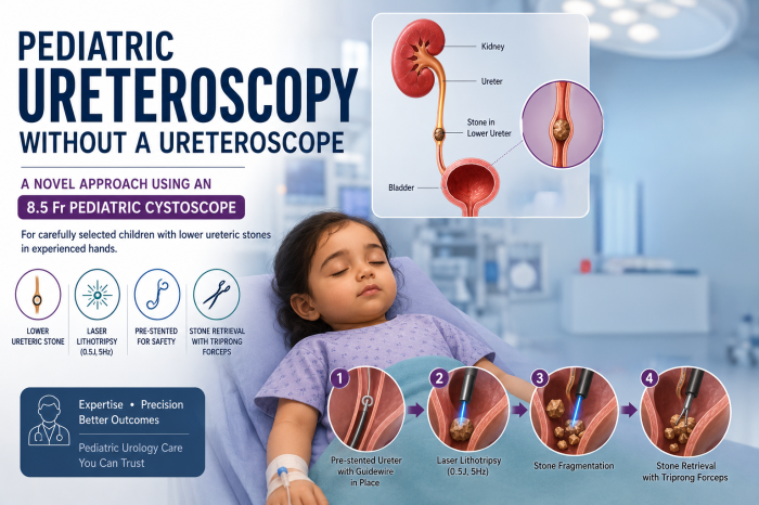
In a recently demonstrated case, Dr. Prabhu, Consultant Pediatric Urologist, successfully managed a lower ureteric stone in a 2-year-old child using an 8.5 Fr pediatric cystoscope instead of a dedicated pediatric ureteroscope. The case highlights how, in carefully selected children and in experienced hands, a pediatric cystoscope can be considered as an alternative for endoscopic stone management.
Why Are Ureteric Stones More Challenging in Young Children?
Children have much smaller urinary tracts than adults, making endoscopic procedures technically demanding. The ureter is narrow and delicate, requiring miniature instruments and meticulous handling to minimize the risk of injury.
Because of these anatomical differences, pediatric stone surgery requires not only specialized equipment but also significant experience in pediatric endourology.
A Clinical Case: Lower Ureteric Stone in a 2-Year-Old Child
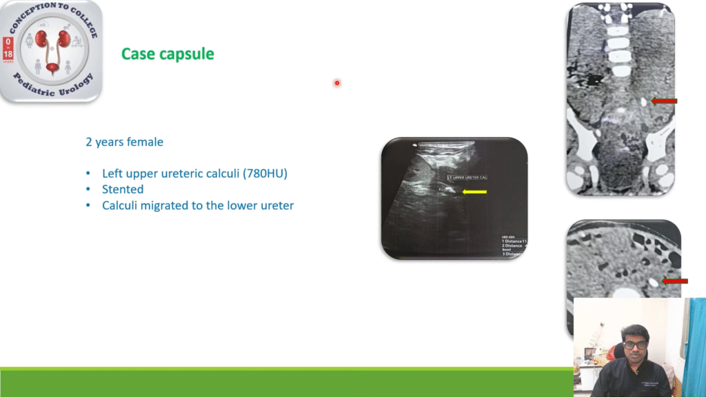
The child presented with left-sided abdominal pain, and imaging with ultrasound and CT scan confirmed a left upper ureteric stone.
Instead of immediate surgery, conservative management was initially chosen. Over the next two weeks, the stone migrated naturally into the lower ureter, making endoscopic removal more feasible.
Rather than using a dedicated pediatric ureteroscope, Dr. Prabhu performed the procedure with an 8.5 Fr pediatric cystoscope, demonstrating an innovative approach for carefully selected pediatric patients.
Step-by-Step Overview of the Procedure
The surgery was performed under general anesthesia using a structured endoscopic technique.
Patient Positioning
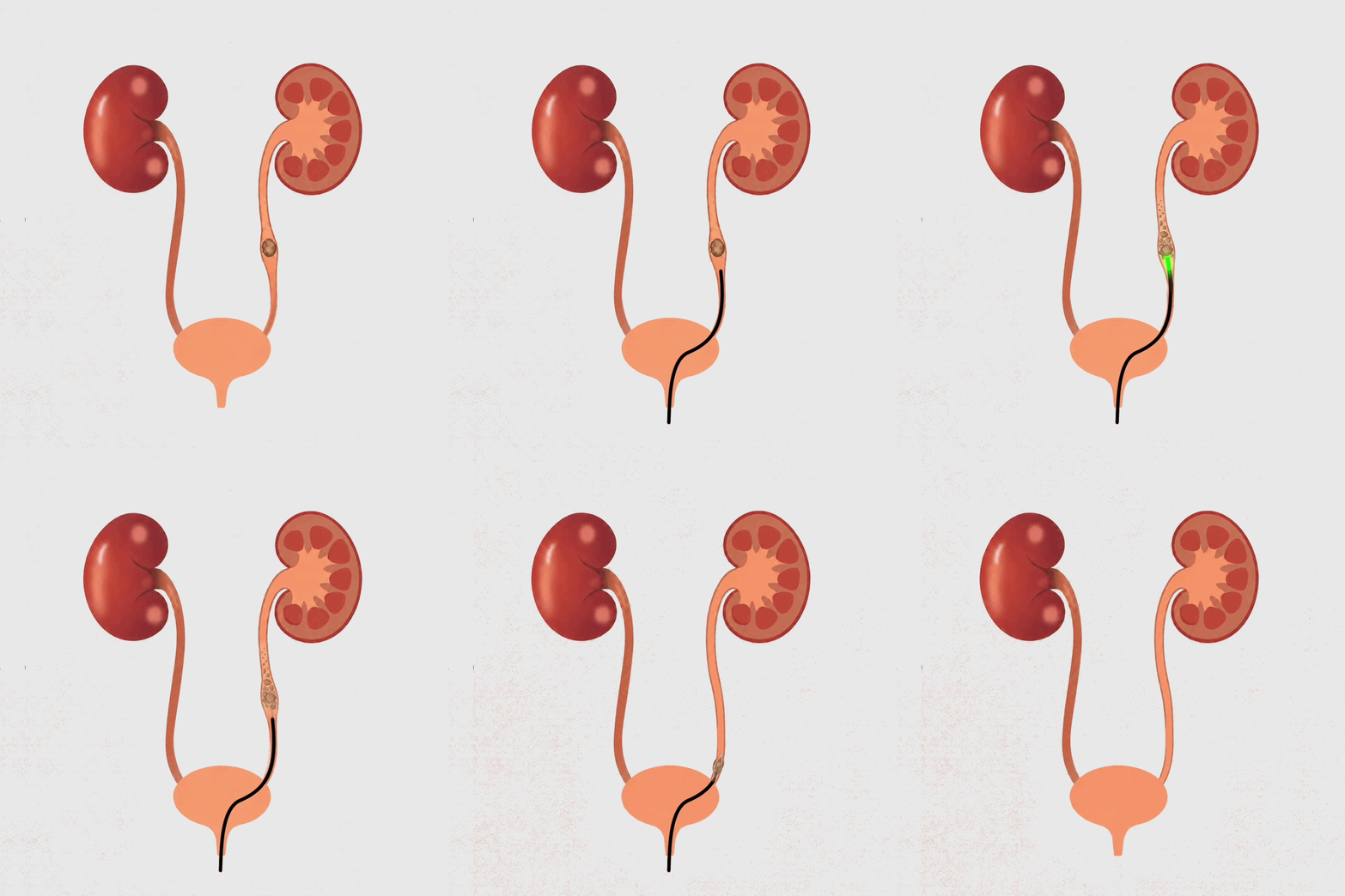
The child was placed in the lithotomy position, and bladder drainage was established to provide continuous venting and maintain clear visualization throughout the procedure.
Guidewire Placement
Before advancing the cystoscope, a guidewire was inserted into the ureter. According to Dr. Prabhu, this provides a safer pathway for the instruments, stabilizes the ureter, and helps reduce stone movement during laser fragmentation.
Pre-stenting
Dr. Prabhu also emphasizes that children selected for this technique are pre-stented, helping facilitate safer access to the ureter before the procedure begins.
Laser Lithotripsy
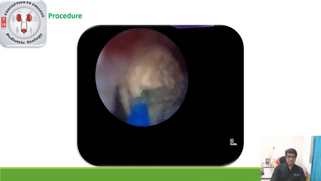
Once the stone was visualized, laser lithotripsy was performed using low-energy settings appropriate for pediatric patients: 0.5 Joules, 5 Hz Approximately, 2.5 Watts. The laser was aimed precisely at the center of the stone to achieve effective fragmentation while minimizing injury to the surrounding ureter.
Stone Retrieval
Following fragmentation, the stone fragments were removed using triprong grasping forceps, completing the procedure.
Why Was an 8.5 Fr Pediatric Cystoscope Used?
Dr. Prabhu explains several practical reasons why an 8.5 Fr pediatric cystoscope was suitable in this carefully selected case.
Readily Available Equipment
Unlike dedicated pediatric ureteroscopes, pediatric cystoscopes are commonly available in pediatric surgical units, making them a practical option when a ureteroscope is unavailable.
Excellent Endoscopic Visualization
The cystoscope provides excellent visualization, allowing accurate identification of the stone and precise laser targeting.
Larger Working Channel
Its larger working channel makes it easier to manipulate the laser fiber and triprong grasping forceps during the procedure.
Easier Maneuverability
With a length of approximately 15 cm, the pediatric cystoscope is easier to maneuver within the shorter ureters of selected young children.
Important Considerations Before Using This Technique
Although this case was successfully managed, Dr. Prabhu stresses that this approach should not replace conventional pediatric ureteroscopy.
Several important factors must be considered before attempting this technique.
Surgical Expertise Is Essential
This procedure should only be performed by surgeons experienced in pediatric endourology. Safe advancement of the cystoscope within the ureter requires careful technique and familiarity with pediatric anatomy.
Not Suitable for Every Child
This approach is feasible only in a small proportion of young children, particularly those with a ureter length of approximately 15 cm.
Stone Location Matters
According to Dr. Prabhu, the technique is most suitable for lower ureteric stones, where access with the pediatric cystoscope is achievable.
When Can This Approach Be Considered?
Based on the operative experience shared by Dr. Prabhu, an 8.5 Fr pediatric cystoscope may be considered when:
- A dedicated pediatric ureteroscope is unavailable.
- The child has been carefully selected and appropriately pre-stented.
- The ureter length is suitable for the procedure.
- The stone is located in the lower ureter.
- The surgery is performed by an experienced pediatric urologist.
Every child should be evaluated individually, and treatment decisions should always be based on the patient’s anatomy and clinical condition.
This case illustrates that innovative solutions can sometimes expand the options available for pediatric stone surgery. In experienced hands and with careful patient selection, an 8.5 Fr pediatric cystoscope may be considered as an alternative to a dedicated pediatric ureteroscope in selected children with lower ureteric stones.
However, as emphasized by Dr. Prabhu, the success of this technique depends on multiple factors, including patient selection, ureter length, stone location, pre-stenting, and surgical expertise.